Catalogue of the Botanical Art Collection at the Hunt Institute
Scroll down after clicking "Search" to view results.
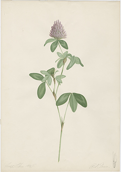
Red clover, Trifolium pratense Linnaeus, Fabaceae alt. Leguminosae
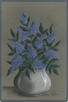
Syringa Linnaeus, Oleaceae
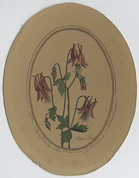
Aquilegia canadensis Linnaeus, Ranunculaceae
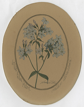
Phlox divaricata Linnaeus, Polemoniaceae
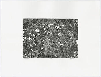
Quercus veluntina Lamark, Fagaceae
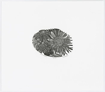
Echinaceae purpurea (Linnaeus) Moench; Helianthus annuus Linnaeus, Asteraceae alt. Compositae
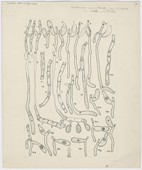
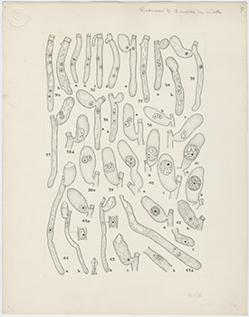
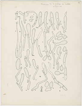
Helicogloea lagerheimi Pat., Auriculariaceae
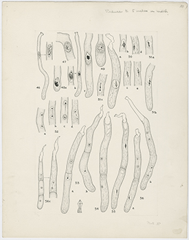
Helicogloea lagerheimi Pat., Auriculariaceae
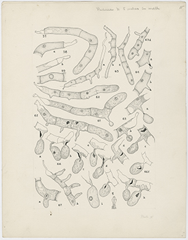
Helicogloea lagerheimi Pat., Auriculariaceae
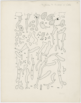
Helicogloea lagerheimi Pat., Auriculariaceae
Helicogloea lagerheimi Pat., Auriculariaceae
Helicogloea lagerheimi Pat., Auriculariaceae
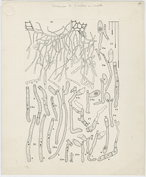
Helicogloea pinicola Bourdot & Galzin; H. pinicola f. alniviridis; Helicogloea graminicola Bresadola; Helicogloea intermedia Linder; Helicogloea caroliniana Coker; Helicogloea lagerheimi Patouillard ; Helicogloea lagerheimi Patouillard, Auriculariaceae
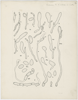
Helicogloea pinicola Bourdot & Galzin; H. pinicola f. alniviridis; Helicogloea graminicola Bresadola; Helicogloea intermedia Linder; Helicogloea caroliniana Coker; Helicogloea lagerheimi Patouillard ; Helicogloea lagerheimi Patouillard, Auriculariaceae

Acorus calamus Linnaeus, Acoraceae

Alpinia galanga Willdenow, Zingiberaceae

Allium tricoccum Aiton, Liliaceae

Alpinia officinarum Hance, Zingiberaceae

Angelica venenosa (J. Greenway) Fernald, Apiaceae

Bixa orellana Linnaeus, Bixaceae

Celtis occidentalis Linnaeus, Ulmaceae

Collinsonia canadensis Linnaeus, Labiatae, Lamiaceae

Chenopodium ambrosioides Linnaeus, Chenopodiaceae

Cryptotaenia canadensis (Linnaeus) de Candolle, Apiaceae

Cunila origanoides (Linnaeus) Britton, Labiatae alt. Lamiaceae